Project Description:
Epilepsy affects 2.2 million Americans and 65 million people worldwide. Seizures have the potential to kill neurons and cause brain injury, leading to reorganization of neural circuits, and progressively more severe epilepsy. Our goal is to quantify neural injury caused by seizures, to provide information to physicians that will assist in selecting optimal treatments for epileptic patients. This quantification is difficult to carry out with current histopathology methods due to inability to compare pre- and post-seizure neural tissue.
We plan to overcome these limitations by carrying out Optical Coherence Microscopy (OCM) imaging of an in vitro organotypic hippocampal culture model of epilepsy before, during, and after seizures. OCM is an emerging optical imaging technique that enables micron-scale, cross-sectional, and three-dimensional (3D) imaging of biological tissues in situ and in real-time. OCM functions as a type of “optical biopsy,” imaging tissue microstructure with resolutions approaching that of standard histopathology, but without the need to remove and process tissue specimens. We will validate the use of OCM to non-invasively quantify the numbers of neurons in organotypic hippocampal cultures, and then, correlate changes in neural numbers measured with OCM to duration and type of spontaneous seizures.
Results of this project will lead to a better understanding of the relationship between seizures and death of neurons, potentially leading to improved treatments for patients with epilepsy.
Project Year:
2013
Team Leaders:
Chao Zhou, Ph.D. (Electrical & Computer Engineering and Bioengineering)
Yevgeny Berdichevsky, Ph.D. (Electrical & Computer Engineering and Bioengineering)
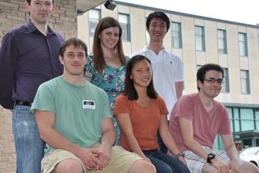
Graduate Students:
Fengqiang Li
Yu Song
Undergraduate Students:
William Cogguillo
Jonah Kohen
Nicole Pirozzi
Anna Sternberg


